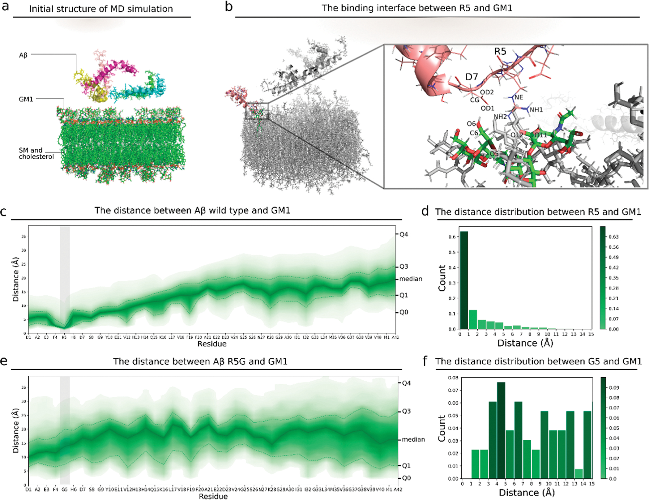
Figure 2.

Interaction between the wild type, mutant Aβ, and GM1 liposome. (a) Initial structure for the MD simulation of the GM1 membrane and 5 Aβ monomers. (b) 3D view of the atoms at the interacting interface between R5 and GM1. (c) Distances between atoms of each residue in wild type Aβ and atoms of GM1 in the membrane. The upper area edge, the upper dashed line, the solid line, the lower dashed line, and the lower area edge are the 100, 75, 50, 25, and 0 percentiles, respectively, for the distance between each residue and GM1. The color corresponds to the percentile. The gray vertical bar indicates the region of the fifth residue. (d) Histogram of the minimum distances between R5 in Aβ and GM1 in the membrane of all steps in the MD simulation. (e) Distances between the Aβ mutant R5G and GM1. (f) Histogram of the minimum distances between G5 and GM1. G5 is the fifth residue in the Aβ mutant R5G.
