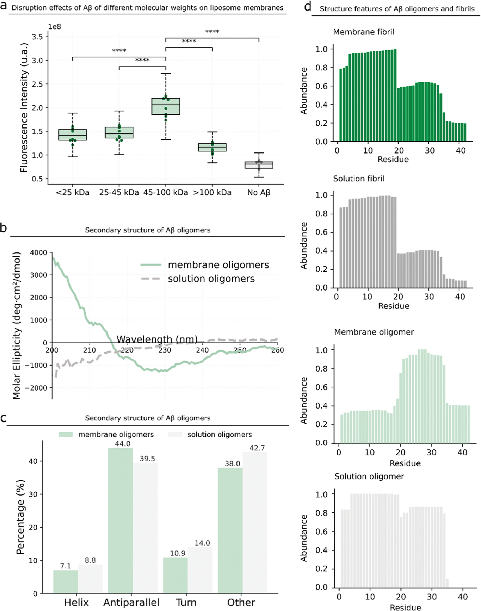
Figure 3.

Structure characterization of membrane Aβ oligomers. (a) We divide the membrane Aβ oligomers into four samples according to the molecular weights and purify them from gels. We incubate the four samples at the same concentration with the Ca2+-encapsulated liposome and determine the fluorescence intensity. P-value: NS (0.05 < p ≤ 1), * (0.01 < p ≤ 0.05), ** (0.001 < p ≤ 0.01), *** (0.0001 < p ≤ 0.001), and **** (p ≤ 0.0001). (b,c) Secondary structure of the Aβ oligomers formed in the presence and absence of GM1 liposomes. (d) Residue abundance of Aβ fibrils formed in the presence and absence of GM1 liposomes and the residue abundance of Aβ oligomers formed on the membrane and in the absence of GM1 liposomes.
