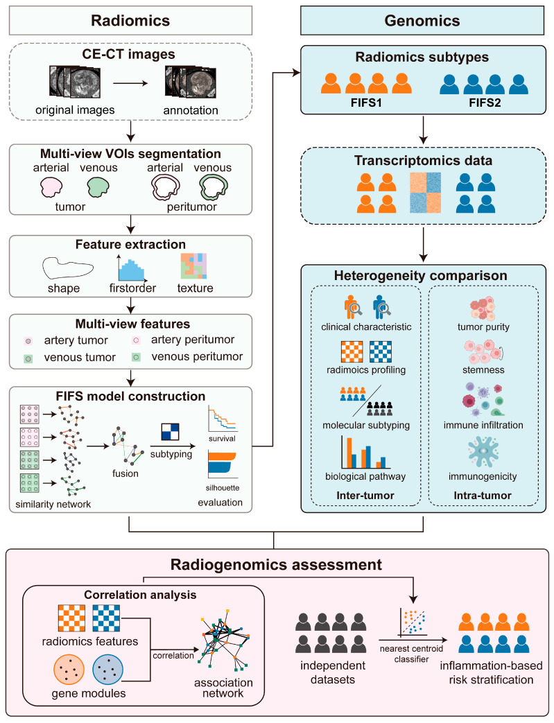
Figure 1.
Schematic diagram of the study. We divide the workflow into three main steps. First, we segment CE-CT images to acquire multi-view VOIs (tumor and peritumoral regions in the arterial and venous phase, respectively), followed by extracting multi-view imaging features. Next, the FIFS model is developed for feature fusion and radiomics subtype identification. Based on the corresponding gene expression profiles and imaging features, we compare inter- and intra-tumor heterogeneity between subtypes. Finally, radiogenomics association is demonstrated by integrating feature–pathway network analysis and validated by inflammation-based risk stratification in two independent cohorts.

