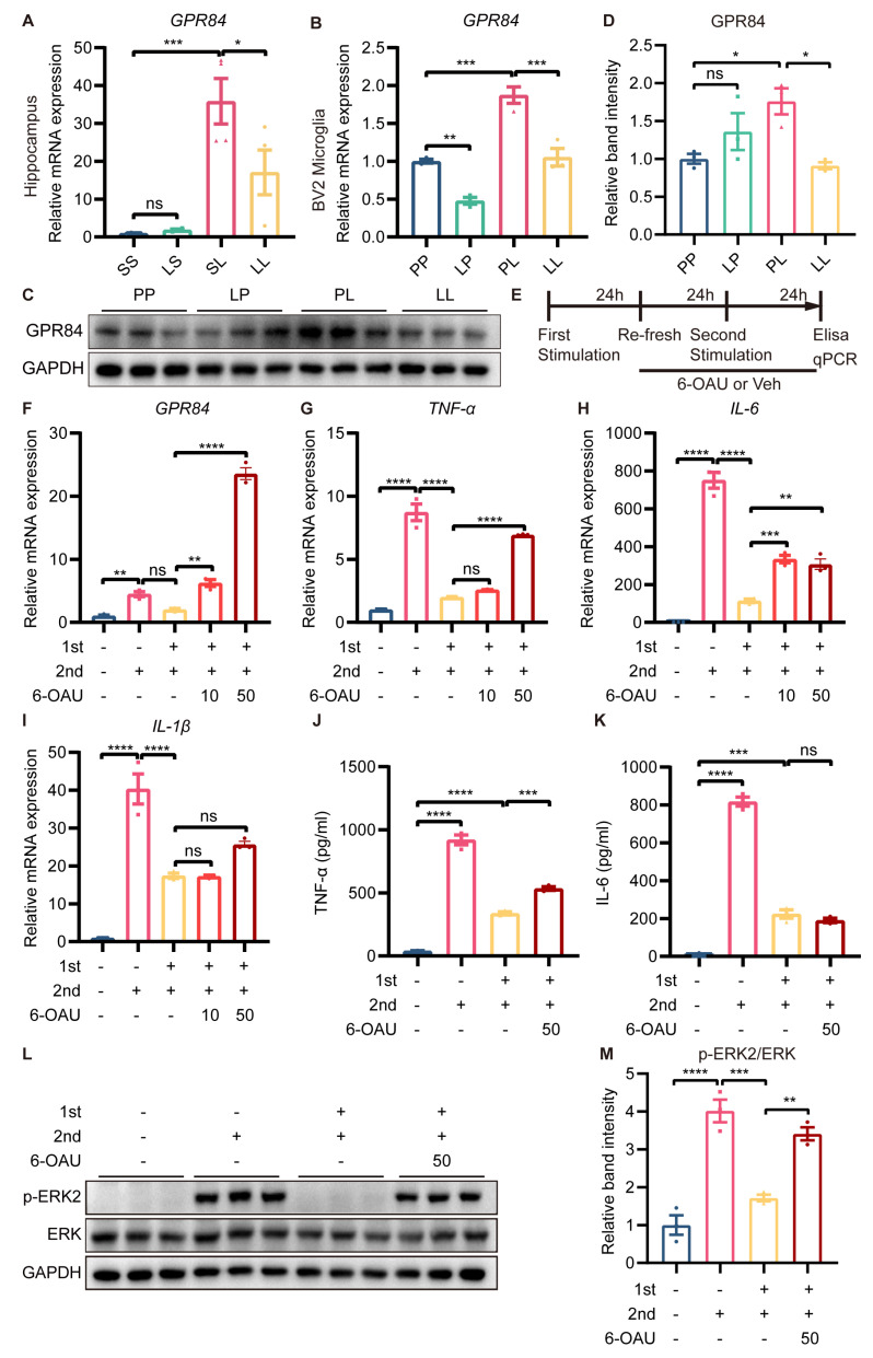
Figure 7.
Effect of GPR84 activation on LPS−induced microglial tolerance−like state. GPR84 mRNA expression in the mouse hippocampus (n = 4) (A) and in BV2 microglia (n = 3) (B). (C) Representative Western blots of GPR84 and the normalization control GAPDH in BV2 microglia. (D) Statistical analysis showed that the expression of GPR84 protein in BV2 microglia was increased in the PL group compared to the PP group, but decreased in the LL group compared to the PL group (n = 3). (E) Schematic diagram of the 6−OAU treatment experiment. “+” referred to LPS treatment, “-“ referred to saline/vehicle treatment, “10” referred to 10 μM, and “50” referred to 50 μM. (F–I) mRNA expression levels of GPR84, TNFα, IL−6 and IL−1β in BV2 microglia. (J,K) Protein levels of TNFα and IL-6 in the cell culture supernatant (n = 3). (L) Representative Western blots of p−ERK2 and ERK in BV2 microglia; the level of each protein was normalized to the GAPDH level (n = 3). (M) Statistical analysis showed that the phosphorylation of the ERK protein in BV2 microglia was increased in the LL + 6OAU group compared to the LL + vehicle group. The data were presented as the mean ± SEM; one−way ANOVA, ns indicated no significance, * p < 0.05; ** p < 0.01; *** p < 0.001; **** p < 0.0001.

