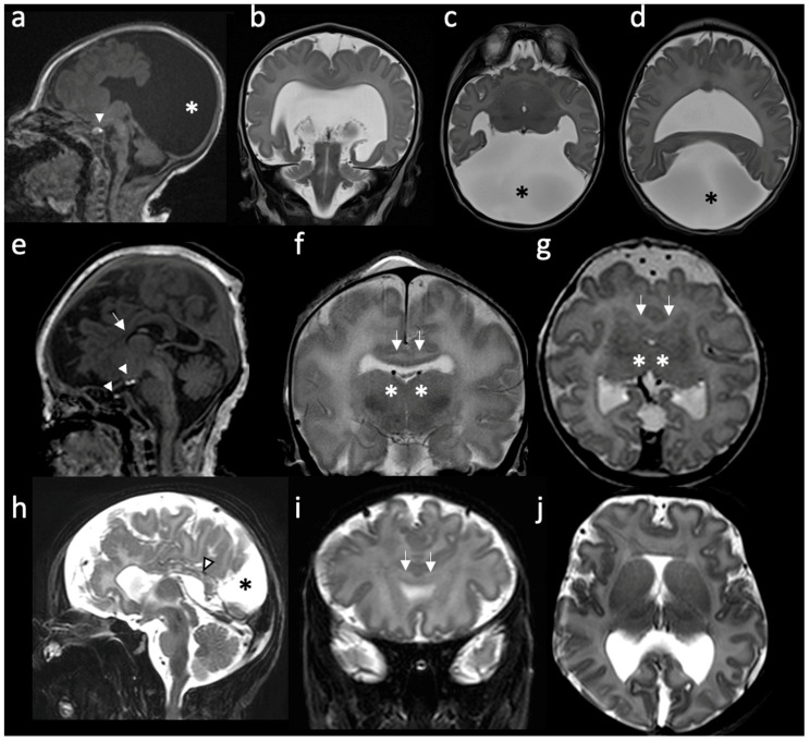
Figure 1.
Radiologic features of holoprosencephalies: (a–d) Radiologic features of alobar holoprosencephaly, 2 days old. (a) Sagittal T1-weighted image shows hypodevelopment of the parietal lobes with absence of identifiable occipital lobes and corpus callosum. The bright T1 spot of the neurohypophysis is preserved and well placed (white arrowhead). (b) Coronal T2-weighted image shows the frontal lobes fused across the midline and a large supratentorial monoventricle. (c,d) Axial T2-weighted images show fused thalami and the monoventricle communicating with a large dorsal cyst (asterisk), noting the absence of the septum pellucidum and rudimentary formation of the temporal horns. (e–g) Radiologic features of semilobar holoprosencephaly, 3 days old. (e) Sagittal T1-weighted image shows hypoplastic frontal lobes, absence of anterior corpus callosum (white arrow) and abnormal finding of partial ectopic neural hypophysis associated with residual hyperintense signal seen within the sella (white arrowheads). (f) Coronal T2-weighted image shows fusion of the frontal lobes (white arrows) and partial fusion of thalami (asterisk). (g) Axial T2-weighted image shows that the division of the ventricles is only seen posteriorly. Septum pellucidum is absent. (h–j) Radiologic features of middle interhemispheric variant holoprosencephaly, 15 days old. (h) Sagittal T2-weighted image shows absent body of the corpus callosum but with the presence of the splenium (white arrowhead). Dorsal interhemispheric cyst is present (black asterisk). (i) Coronal T2-weighted image shows fusion of the frontal lobes across the midline (white arrows). Note that the degree of fusion is less extensive than the one seen in the semilobar HPE. (j) Axial T2-weighted image shows fused frontal lobes, absent septum pellucidi and the interhemispheric cyst in the occipital region.

