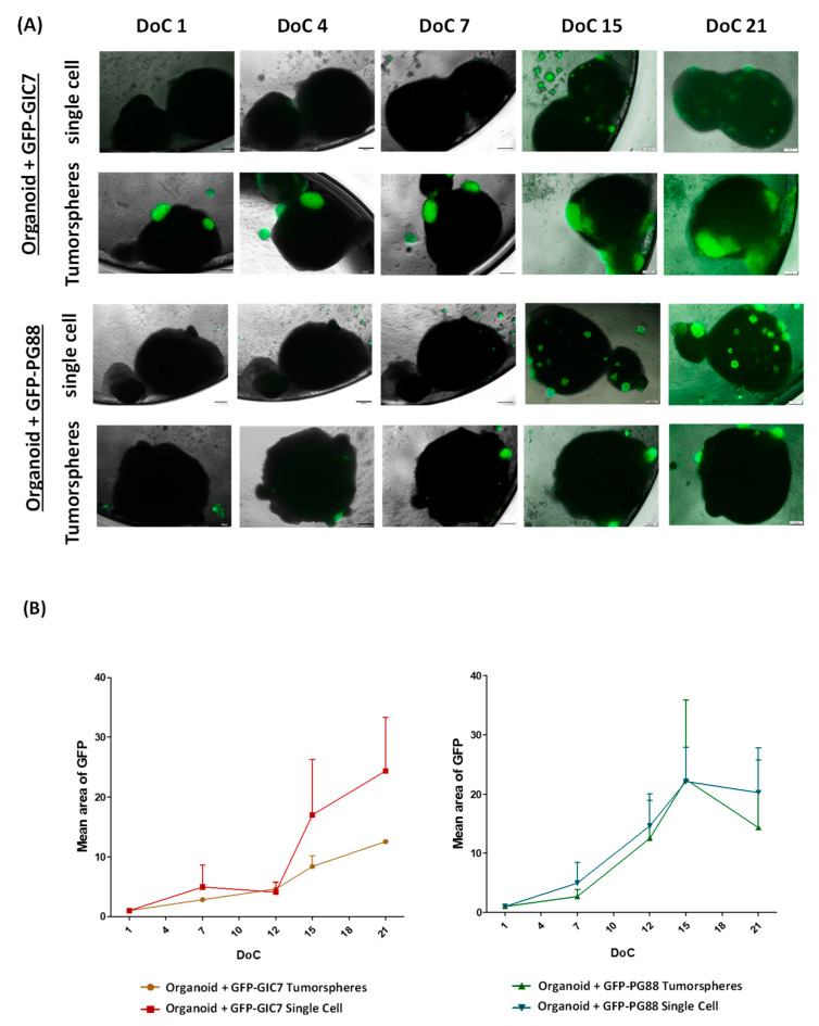
Figure 4.
Study of the tumor engraftment effectiveness. (A) Merge of fluorescence and phase-contrast images of GFP-GIC-organoid co-cultures at days 1, 4, 7, 15, and 21 after the engraftment (DoC). Both GFP-GIC7 (top panels) and GFP-PG88 (bottom panels), seeded as tumorspheres or as a single cell, as indicated, achieve infiltration of the organoid generating a viable tumor. Both GFP-GICs grew around and inside the organoid. Images at 4× obtained with Microscope Olympus IX51 are shown. Scale bar 200 μm. (B) Semi-quantitative analyses of tumor growth expressed as the mean of tumor area (GFP positive area), concerning the total organoid, evaluating GFP fluorescence of GFP-GIC7 (left) and GFP-PG88 (right) seeded as tumorspheres or single cells. To normalize the data and homogenize the different co-cultures, the initial GFP fluorescence on DoC 1 was used to evaluate each well and to compare the GFP area. The data are representative of the tumor engraftment effectiveness of both GFP-GICs. An ANOVA test was used to compare the means of 4 different conditions for every DoC and each situation for every DoC vs. the first DoC. DoC = Days of co-culture.

