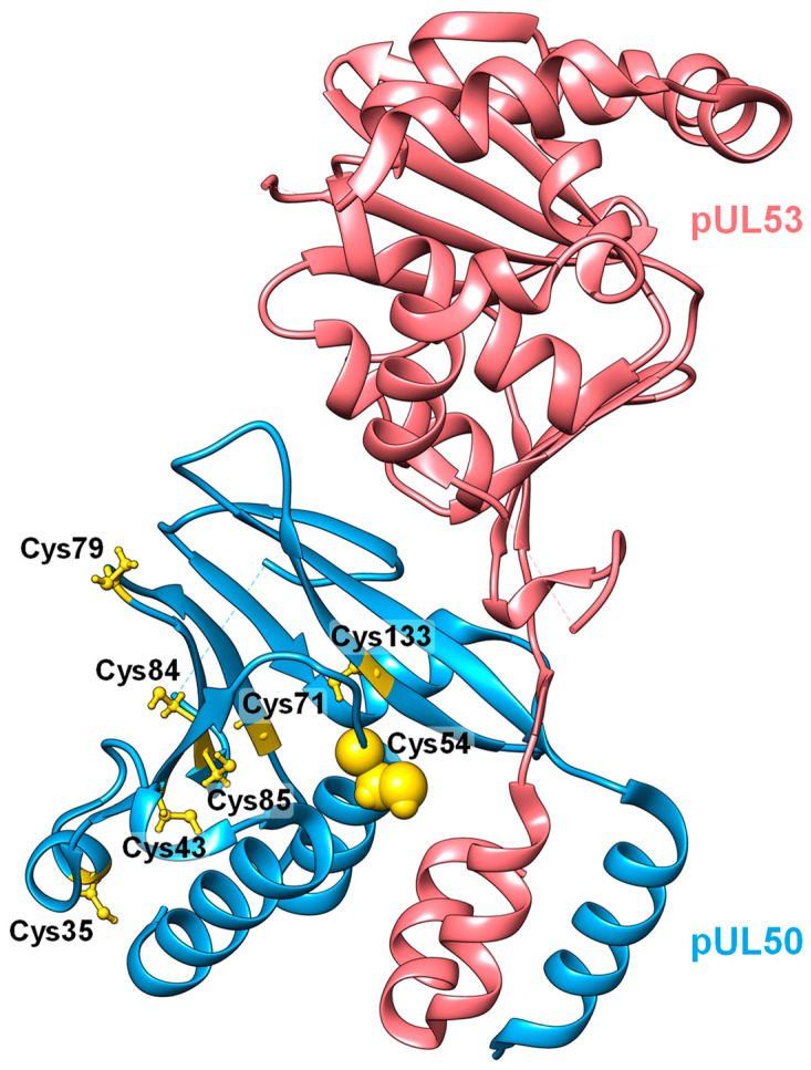
Figure 3.
Protein complex structure of pUL53 and pUL50 from HCMV (PDB ID code: 5D5N [43]). HCMV pUL53 (red) interacts with pUL50 (blue) via a hook-like element that binds into an α-helical groove on pUL50. Cysteine residues in pUL50 are depicted with a yellow ball-and-stick representation, while Cys54, which is the only one of the eight cysteines located at the interaction interface with pUL53, is highlighted with spheres. Protein visualization was performed with UCSF Chimera [67] (version 1.16).

