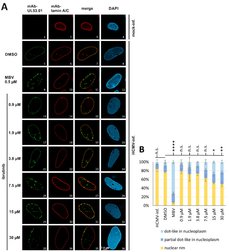
Figure 11.
Inhibitory impact of hit compound ibrutinib on the localization of HCMV pUL53. (A) HFFs were infected with HCMV AD169 (MOI 0.1) and immediately treated with the warhead compound. Ibrutinib was applied at the indicated concentrations, and the determination was performed in biological triplicates. At 5 d p.i., cells were fixed, used for IF staining with the indicated antibodies and analyzed for intranuclear localization by confocal imaging. DAPI counterstaining represented the nuclear morphologies of the respective cells. Additional single channel images in grayscale, allowing easier comparison of signal patterns, are presented in Figure S5. For raw data, see https://doi.org/10.5281/zenodo.7794233 accessed on 3 April 2023. (B) Quantitation of this IF analysis was achieved by counting 50 cells per biological triplicate. Three patterns of pUL53 localization were distinguished, i.e., normal nuclear rim (yellow), partial dot-like intranucleoplasmic aggregation (dark blue) and predominant dot-like aggregation (light blue). Mean values ± SDs are given. Statistical analysis was performed using ordinary one-way ANOVA and post hoc Tukey testing on combined dot-like pUL53 localization values of DMSO in relation to the analyzed ibrutinib concentrations and MBV; * p ≤ 0.05, ** p ≤ 0.01, **** p ≤ 0.0001; n.s., not significant.

