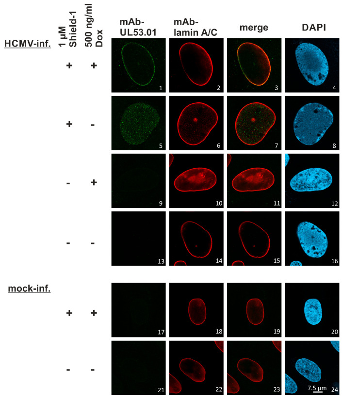
Figure 14.
Shield-1- and Dox-controlled expression of viral core NEC proteins during HCMV ∆UL50-ΣUL53 infection. HFF-UL50 cells were infected with HCMV ∆UL50-ΣUL53 at MOI of 0.3. To analyze the experimental controllability of viral core NEC expression and intranuclear localization, 1 µM of Shield-1 and/or 500 ng/mL of Dox (refreshed every second day) were added as indicated. Uninfected cells served as a negative control (mock). At 6 d p.i., cells were fixed, used for an immunofluorescence staining by the indicated antibodies and analyzed by confocal imaging. Lamin A/C-specific counterstaining was used as a marker of the nuclear rim, representing the typical localization site of viral pUL50–pUL53 recruitment; DAPI counterstaining was used to monitor the morphologies of cell nuclei. For raw data, see https://doi.org/10.5281/zenodo.7794233 accessed on 3 April 2023.

