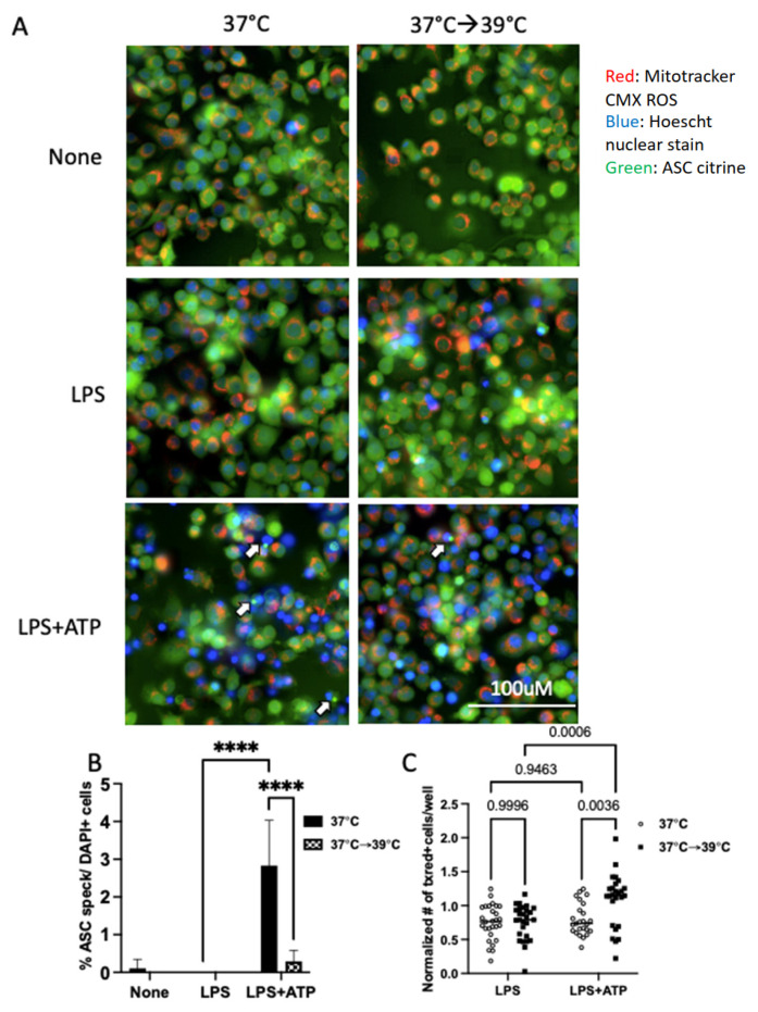
Figure 4.
Mild heat shock inhibits the formation of ASC specks but does not inhibit mitochondrial membrane potential. (A) Representative images of iBMMs expressing ASC-citrine stained with Mitotracker CMX ROS and Hoescht nuclear stain show inhibition of ASC specks (white arrows) and increased numbers of cells with polarized mitochondria in heat-exposed cells. Live cells were imaged using the IXM live cell microscopy system. Images are from a representative experiment. (B) Quantification of ASC specks per DAPI nuclei. Results are a representative experiment of 3 independent experiments, counted manually with >5 (40×) fields/conditions. (C) Quantification of the number of cells/well with respiring mitochondria, normalized to the unstimulated 37 °C control condition, greater than 16 wells/condition/experiment combined over three independent experiments. **** p < 0.0001 as determined by a two-way ANOVA with Tukey’s multicorrection comparison; ns, nonsignificant.

