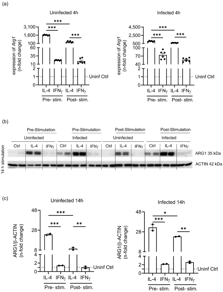
Figure 3.
Priming of macrophages before S.tm infection shows differences in ARG1 regulation. Unpolarised BMDM and pre-stimulated polarised BMDM (2 ng/mL IL-4 or 20 ng/mL IFNγ) were infected with S.tm for 1 h. Unpolarised cells were then stimulated (post-stimulation) with 10 ng/mL IL-4, 100 ng/mL IFNγ or left unstimulated for 4 h or 14 h. ARG1 mRNA and protein levels in pre- and post-stimulation conditions were analysed in uninfected and infected macrophages. (a) Arg1 gene expression depending on stimulation (IL-4 or IFNγ) and infection. Arg1 transcript levels were determined by quantitative real-time PCR and normalized to Hypoxanthine phosphoribosyltransferase (Hprt) mRNA levels using the ΔΔCT method. (b) Western blot analysis of ARG1 and β-ACTIN. (c) Densidometrical quantification of immunoblotting results relative to β-ACTIN expression. Statistical significance was determined by one-way ANOVA with Tukey post hoc test (a + c). * p-value < 0.05; ** p-value < 0.01; *** p-value < 0.001. Representative data from three independent experiments (mean ± SEM) performed in technical duplicates are shown. Data were normalised to the unstimulated, uninfected Ctrl.

