
Figure 3.
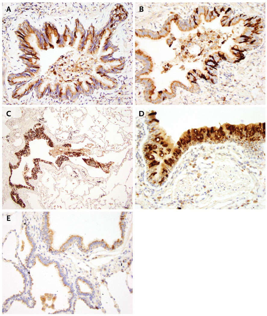
MUC5AC Immunohistochemistry.
Panels A to D show that, by immunohistochemistry, there was overexpression of MUC5AC in the respiratory epithelium of numerous bronchioles in all vaping patients. Panel E shows MUC5AC expression in a nonvaping, former-smoker patient as a control, which has very little MUC5AC expression compared with the vaping patients. Original magnification is as follows: 400× in Panel A (patient 1), Panel B (patient 2), Panel D (patient 4), and Panel E (nonvaping, former-smoker control); and 100× in Panel C (patient 3).