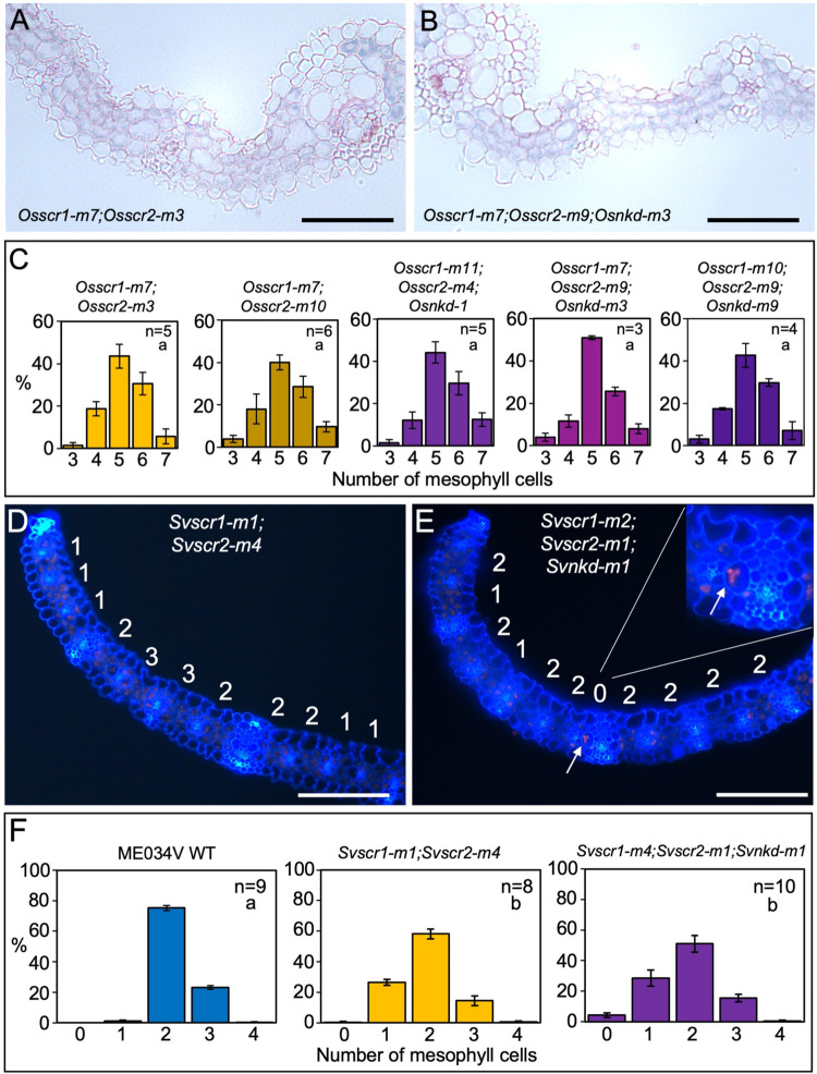
Fig 6. Loss of function nkd mutations induce the formation of fused leaf veins in scr mutants of setaria but not rice.
A-B) Transverse sections of Osscr1-m7;Osscr2-m3 (A) and Osscr1-m7;Osscr2-m9;Osnkd-m3 (B) mutant leaves, taken at the midpoint along the proximal-distal axis of leaf 5. C) Histograms summarizing the mean number of mesophyll cells separating veins in two independent Osscr1;Osscr2 (yellow) and three independent Osscr1;Osscr2;Osnkd (purple) mutant lines. Error bars are standard error of the mean (raw data in S1 Table). Sample sizes (n =) are biological replicates. D-E) Transverse sections of Svscr1-m1;Svscr2-m4 (D) and Svscr1-m2;Svscr2-m1;Svnkd-m1 (E) mutant leaves, taken at the midpoint along the proximal-distal axis of leaf 4, imaged under UV illumination. The number of mesophyll cells separating veins is displayed above each region. In (E) white arrows and the inset show an example of a fused vein with no separating mesophyll cells. F) Histograms summarizing the mean number of mesophyll cells separating veins in wild-type (WT) ME034V (blue), Svscr1-m1;Svscr2-m4 (yellow) and Svscr1-m4;Svscr2-m1;Svnkd-m1 (purple) mutants. Error bars are standard error of the mean. Sample sizes (n =) are biological replicates and letters in the top right corner (beneath sample sizes) of each plot indicate statistically different groups (P≤0.05, one-way ANOVA and Tukey’s HSD) calculated using the mean number of mesophyll cells in each genotype (raw data in S1 Table). Scale bars: 100 μm.

