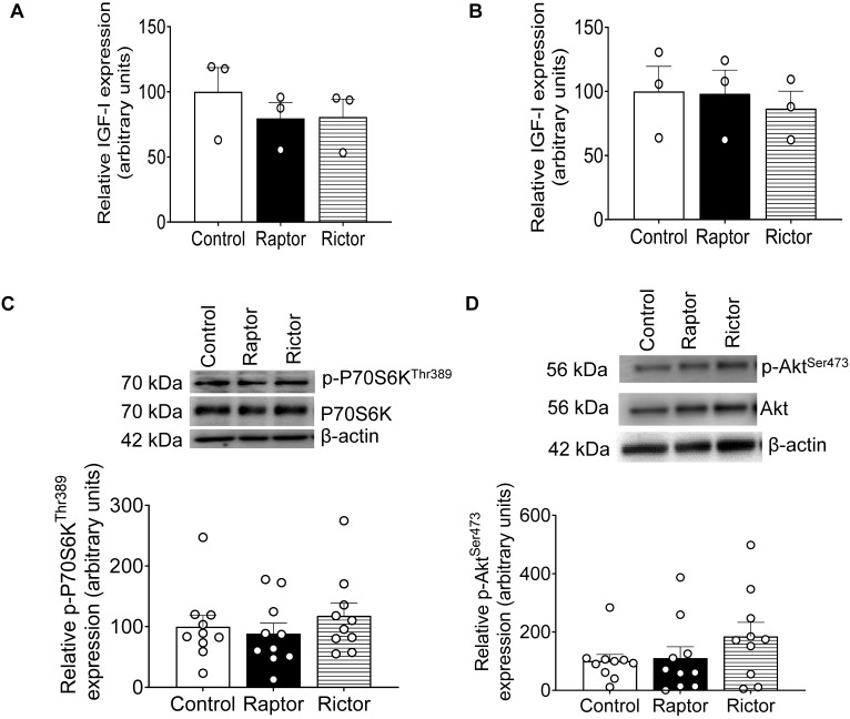
Figure 8.
ELISA analysis of IGF-1 secretion before and after treatment of HepG2 cells with CM from PHT cells with the control, RAPTOR, and RICTOR siRNA and densitometric analysis of mTORC1 and mTORC2 functional readouts from cell lysates of HepG2 treated with CM from PHT cells with the control, RAPTOR, and RICTOR siRNA. IGF-1 secretion was determined in CM from PHT cells with the control, RAPTOR, and RICTOR siRNA before treatment of HepG2 cells (A) and CM from HepG2 cells after treatment with CM from PHT cells with the control, RAPTOR, and RICTOR (B). Equal aliquots (50 μL) of CM were analyzed with an IGF-1 ELISA kit. A representative Western blot of HepG2 cell lysates (35 μg protein) displaying p-P70S6K Thr389 (C) and p-Akt Ser473 (D) in HepG2 cells treated with CM from PHT cells with the control, RAPTOR, or RICTOR siRNA. Samples were normalized to the control. The data were represented as Mean + SEM in bar figures and analyzed with a one-way ANOVA and Bonferroni Multiple Comparison tests; p < 0.05 was considered significant.

