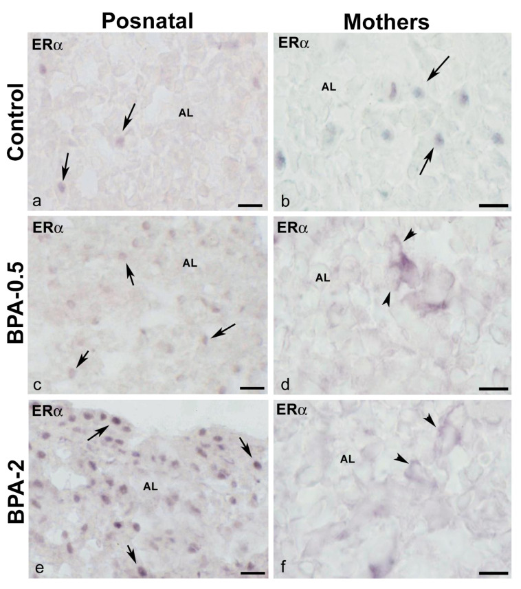
Figure 7.
ERα immunoreactivity in offspring and the mother’s pituitary gland. Representative images of the pituitary gland in offspring at the first week of postnatal life (a,c,e) and mothers after weaning/lactation (b,d,f) showing the presence of ERα-ir cells in the anterior lobe in the three experimental groups. The nuclear location of ERα immunoreactivity in offspring and control mothers is indicated with arrows, while the cytosolic location in BPA-treated mothers is indicated with arrowheads. AL: anterior lobe. Scale bars: (a,c,e): 25 µm and (b,d,f): 20 µm.

