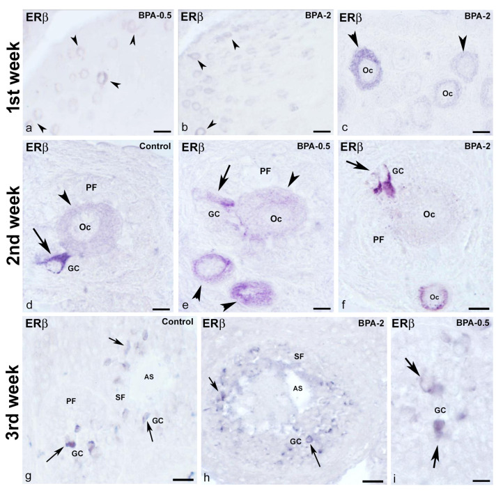
Figure 12.
ERβ immunoreactivity in offspring ovaries. Representative images of the ovary in the three experimental groups in the three first weeks of postnatal life showing the presence of ERβ-ir in oocytes (arrowheads) from primordial follicles in the first week (a–c), in oocytes (arrowheads) from primordial and primary follicles as well as in granulosa cells (arrows) from primary follicles in the second week (d–f) and granulosa cells (arrows) from primary and secondary follicles in the third week (g–i). AS: antral space; GC: granulosa cells; Oc: oocyte; PF: primary follicle; SF: secondary follicle. Scale bars: (a,b): 40 µm; (c,f): 20 µm; (d,e,i): 10 µm; (g): 45 µm; (h): 36 µm.

