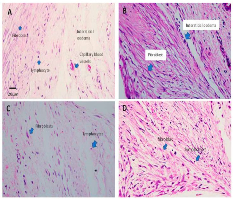
Figure 5.
Histopathological findings of all study groups at post-op day 21. (A) Group A shows dense fibroblasts, which are characterised by spindled-shaped cells that produce collagen, forming scar tissue. The lymphocyte infiltrates are scattered within the tissue. The interstitial oedema is obvious. (B) Group B shows a strong cellular response due to the increased proliferation of fibroblasts (bluish spindled nuclei) and collagen fibres (pink eosinophilic stain fibres). Note the increase in the number of capillary blood vessels lined by endothelial cells. (C) Group C shows an increased cellular response with a proliferation of fibroblasts. The fibroblasts are densely packed and plumped, producing collagen fibres. (D) Group D shows an increased cellular response with a proliferation of fibroblasts. The fibroblasts are densely packed and plumped, producing collagen fibres. Lymphocytes are scattered but reduce in number. Blue arrows in all images pointing the labelling structures (H&E staining, 400× magnification).

