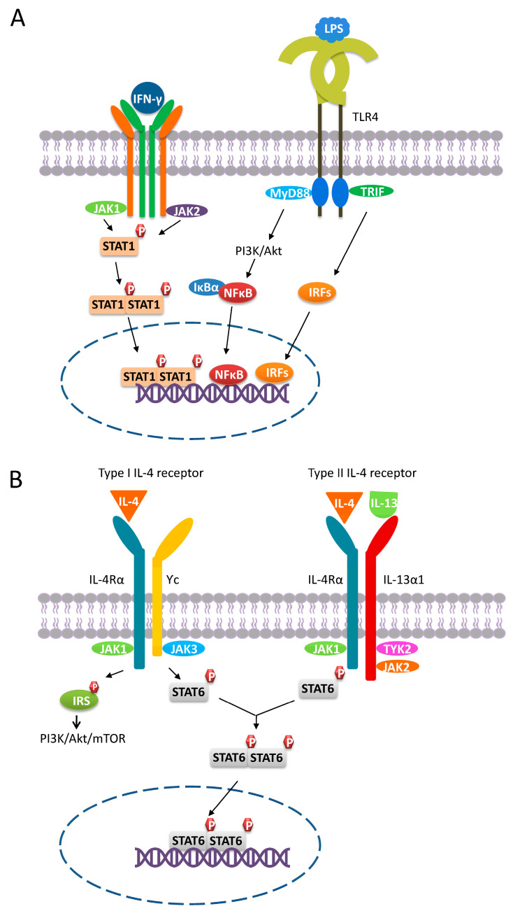
Figure 1.
M1 and M2a polarization. (A) M1 macrophages polarize through signaling pathways mediated by STAT1, PI3K/Akt, NFκB, and IRFs. Binding of IFN-γ to its receptor leads to the recruitment of JAK1 and JAK2, inducing the phosphorylation of STAT1, which then dimerizes and translocates into the nucleus. In response to the LPS-TLR4 engagement, PI3K/Akt-, NFκB-, and IRFs-mediated signaling are triggered. (B) M2a macrophages polarize through the PI3K/Akt- and STAT6-mediated signaling pathways. Macrophages express both type I and type II IL-4 receptors. The engagement of the type I/II IL-4 receptor results in the phosphorylation and subsequent dimerization of STAT3. Once activated, STAT3 dimers translocate into the nucleus and trigger corresponding gene expression. In contrast, IRSs can only be activated by the type I IL-4 receptor and do not translocate to the nucleus. Instead, activated IRSs can induce signaling pathways such as PI3K/Akt-mediated signaling.

