Figure 3.
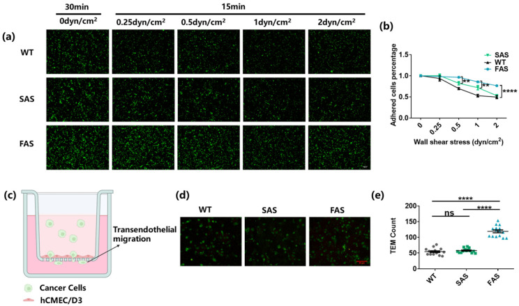
FAS cells exhibit enhanced adhesion to the brain endothelium and BBB transmigration ability. (a,b) FAS cells exhibited enhanced adhesion strength on the endothelium. All groups were labeled with green cell tracker and added into the flow chamber slides to adhere to the brain endothelium for 30 min. Then, 0.25, 0.5, 1, and 2 dyn/cm2 wall shear stress were applied to each slide for 15 min, respectively. The cancer cells remaining on the brain endothelium after each treatment of fluid shear stress were counted using the fluorescence microscope. Two-way ANOVA along with post hoc Tukey test were used to calculate the statistics. Scale bar = 100 μm. n = 3. (c) The illustration of trans-endothelial migration assay (created by biorender.com). (d,e) FAS cells exhibited enhanced BBB transmigration ability. An hCMEC/D6 monolayer was cultured on the top of the insert membrane and cancer cells were added to transmigrate through the monolayer to the lower chamber. The transmigrated cancer cells (marked by the green cell tracker) were imaged and counted using fluorescence microscopy. One-way ANOVA along with post hoc Bonferroni test was performed to analyze the statistics. Scale bar = 50 μm. n = 3. All data are represented by mean ± SEM. (ns: no significance, ** p < 0.01, **** p < 0.0001).

