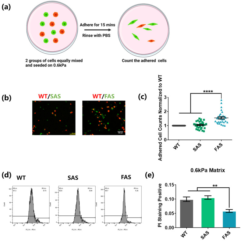
Figure 4.
FAS cells exhibit advantages in cell adhesion and survival within the soft brain environment. (a–c) FAS cells had an enhanced adhesion ability on 0.6 kPa soft matrices. The same number of cancer cells from the WT group (labeled with red cell tracker) was mixed with cancer cells from the FAS group or the SAS group (labeled with green cell tracker) and seeded on the same 0.6 kPa polyacrylamide gels coated with collagen I. After 15 min, these cells were gently washed with PBS. This illustration was created using Biorender.com (a). The remaining cells were imaged and counted under fluorescence microscope. Scale bar = 100 μm. n = 3. (d,e) FAS cells exhibited lower cell apoptosis on soft matrices. All groups were seeded on 0.6 kPa polyacrylamide gels coated with collagen I in low-FBS medium overnight. The dead cells were then marked with PI and tested through flow cytometry. n = 3. All data are represented by mean ± SEM. The statistics among three groups were calculated based on one-way ANOVA with the post hoc Bonferroni test. (** p < 0.01, **** p < 0.0001).

