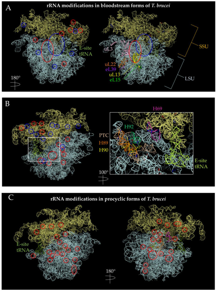
Figure 4.
Positions of the rRNA with hyperpseudouridylations and increased 2′-O-methylations in bloodstream forms (BSF) and procyclic forms (PCF) of T. brucei. PDB 4V8M [104] ribosome structure were used to visualize methylation and pseudouridylation sites using PyMOL [110]. The SSU is indicated by pale yellow, while the LSU is indicated by pale cyan. Methylation sites are circled in red, while pseudouridylation sites are indicated in blue. (A) Methylation and pseudouridylation sites are increased in BSF (+y). There seems to be a cluster of methylation sites in the SSU near the E-site. There is also a cluster of methylation sites near the nascent polypeptide exit tunnel as indicated by uL22 and near uL13 and uL3. uL13 has extra-ribosomal functions in mammals while uL3 has increased expression in certain mammalian tissues [17]. (B) Methylation and pseudouridylation sites in BSF (−y). Left figure shows a view of 180 degrees along the y-axis of (A); right side shows a view of 280 degrees along the y-axis of (A) and a magnification of 60 Å, highlighting helices composing the PTC. Pseudouridylation sites seem to cluster in the SSU, while methylation appears to cluster in the LSU on this side. (C) Increased methylation sites in PCF. Methylation sites appear more diffused in PCF parasites than in BSF forms. The helices composing the PTC are highlighted with the same color pattern as in (B). There are also more methylation sites in PCF than BSF.

