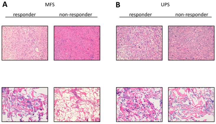
Figure 1.
Representative images of hematoxylin and eosin H&E staining. (A) H&E of MFS responder and non-responder patients’ surgically resected tumor specimen (10× magnification, upper panels) and patient-derived primary culture (10× magnification, lower panels). (B) H&E of UPS responder and non-responder patients’ surgically resected tumor specimen (10×, upper panels) and patient-derived primary culture (10×, lower panels).

