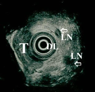
Figure 4.

Corresponding endosonographic image (7.5 MHz) of the same (fig. 3) patient. Hypoechoic mass invading the duodenum. Enlarged hypoechoic regional lymph nodes are also present. Carcinoma of the ampulla of Vater:T3N1. (DL: duodenal lumen, T: tumour mass, LN: metastatic lymph nodes).
