Figure 2.
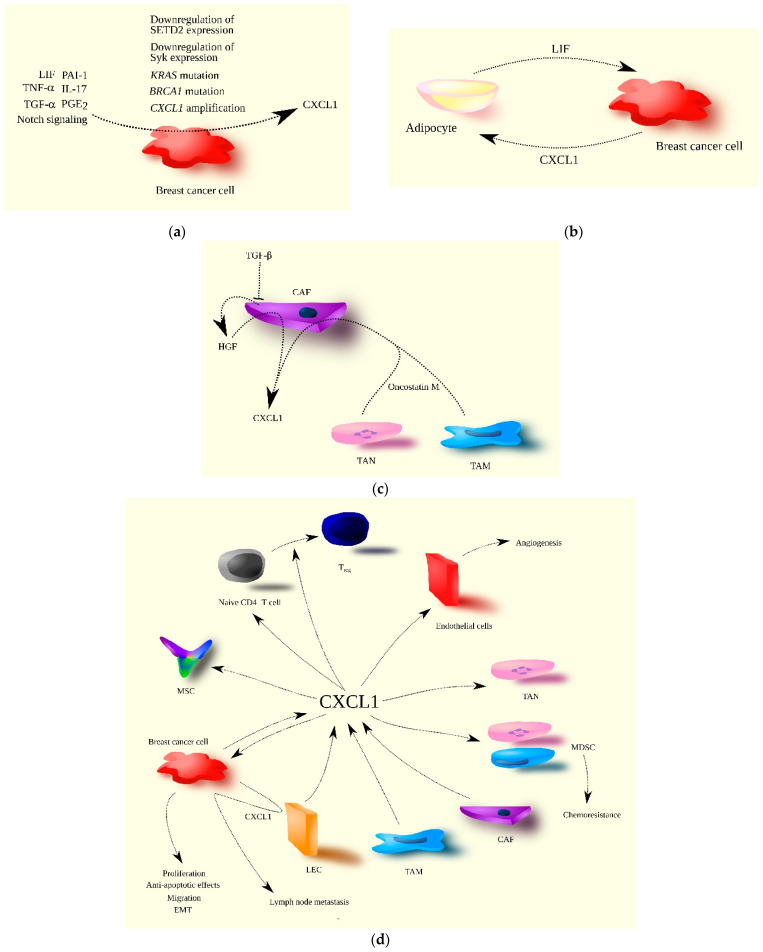
The significance of CXCL1 in breast cancer progression. (a) CXCL1 expression is increased in breast cancer cells by both extracellular and intracellular factors. TNF-α, TGF-α (EGFR ligand), PGE2, IL-17, LIF, PAI-1, and Notch signaling are examples of extracellular factors, while mutations in genes such as KRAS and BRCA1, CXCL1 gene amplification, and the decreased expression of SETD2 and Syk are intracellular factors increasing CXCL1 expression in breast cancer cells. (b) The interaction of breast cancer cells with tumor-associated adipocytes results in increased CXCL1 expression in breast cancer cells due to the secretion of LIF by adipocytes. In turn, CXCL1 increases LIF expression in adipocytes, creating a positive feedback loop between these two types of cells in the breast tumor. (c) In the breast tumor, CXCL1 is secreted by CAF, and its expression in CAF is increased by oncostatin M produced by TAM and TAN. Increased CXCL1 expression in CAF is also due to their loss of responsiveness to TGF-β, resulting in increased HGF production by CAF, which further increases CXCL1 expression. (d) CXCL1 plays multiple roles in breast cancer progression, acting on various types of cells. Source of CXCL1 in breast tumor may be breast cancer cells, CAF, TAM, and LEC. CXCL1 promotes breast cancer cell proliferation, migration, EMT, and anti-apoptotic effects. Breast cancer cells also increase CXCL1 expression in LEC, promoting the migration of breast cancer cells to lymphatic vessels, resulting in lymph node metastasis. CXCL1 also acts on tumor-associated cells. It recruits immune cells such as naïve CD4+ T cells, TAN, MDSC, and MSC to the tumor niche. It can differentiate naïve CD4+ T cells into Treg and facilitate chemoresistance through the recruitment of MDSC. CXCL1 also promotes angiogenesis via its influence on endothelial cells.

