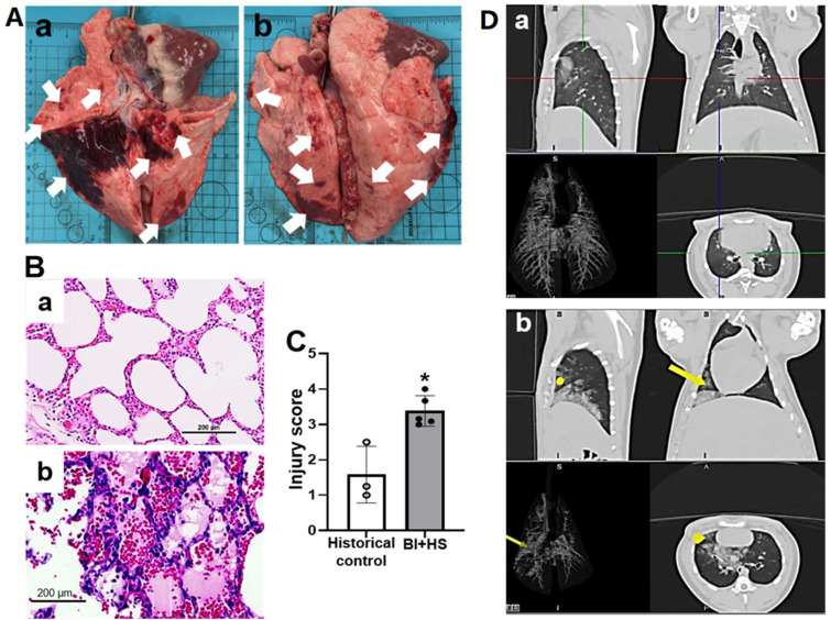
Figure 3.
BI+H-induced acute lung injury (a, anterior; b, posterior) in pigs depicted by gross pathological ((Aa), anterior; (Ab), posterior; white arrows indicating lung injury with diffuse ecchymotic hemorrhage), micropathological alterations of H&E-stained slide [(Ba), historical control group (n = 3); (Bb), B+HS group (n = 5] and histological injury score (C), and CT changes ((Da), pre-injury; (Db), post-injury; yellow dots and yellow arrows depicting opacity in the right accessory and caudal lung lobes, respectively). The data are expressed as mean ± SD. * p < 0.05 vs. historic control. Scale bar = 200 µm.

