Figure 1.
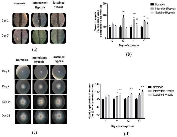
In vitro sustained hypoxia and intermittent hypoxia increase hepatic tumor cell expansion. (a) Representative illustrations of HepG2 cell invasiveness in 2D, assessed by wound healing, before and after 7 days of normoxia, intermittent hypoxia or sustained hypoxia exposure (white scale bar = 600 µm). (b) Wound healing, expressed as a % of repaired area compared with normoxia, of HepG2 cells exposed to 7 days of normoxia, intermittent hypoxia or sustained hypoxia; n = 5 experiments per group with at least 3 wells/experiment. Intermittent hypoxia global effect p < 0.00001, ** p < 0.01 on D4 and D7 for IH vs. N and on D5 and D7 for SH vs. N and *** p < 0.001 for IH vs. N on D5, one-way repeated measures ANOVA. Sustained hypoxia global effect p = 0.006, one-way repeated measures ANOVA and ** p < 0.01 on D5 and on day 7. (c) Representative illustrations of HepG2 spheroid expansion before and after 5, 7, 14 and 21 days of normoxia, intermittent hypoxia or sustained hypoxia exposure (white scale bar = 1000 µm). (d) HepG2 spheroid expansion in response to 21 days of intermittent hypoxia; n = 5 experiments per group (6 to 12 wells/experiment). * p < 0.05, repeated measures ANOVA). Post hoc analysis showed significant differences * p < 0.05 on days 7 and 21 of exposure. HepG2 spheroid expansion in response to 21 days of sustained hypoxia; n = 5 experiments per group (6 to 12 wells/experiment). * p < 0.01, repeated measures ANOVA. Post hoc analysis showed significant differences * p < 0.05 on days 7, 14 and 21 of exposure.

