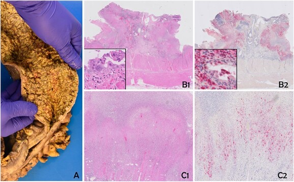
Figure 2.

The specimen shows severe diffuse necrotizing colitis (A). Histology reveals extensive ulcerous colitis with deep-reaching ulcers and mural necrosis (B1 and C1). Multinucleated epithelial cells with eosinophilic inclusions can be discerned (insert B1). Immunohistochemistry for human HSV confirms a widespread infection of the mucosa and the mural part of the colonic wall (B2 and C2). Epithelial cells, as well as stromal cells, are positive (insert B2). B1 and C1: Hematoxylin and Eosin; C1 and C2: Immunohistochemistry for HSV (Chromogen: Fast Red).
