Figure 3.
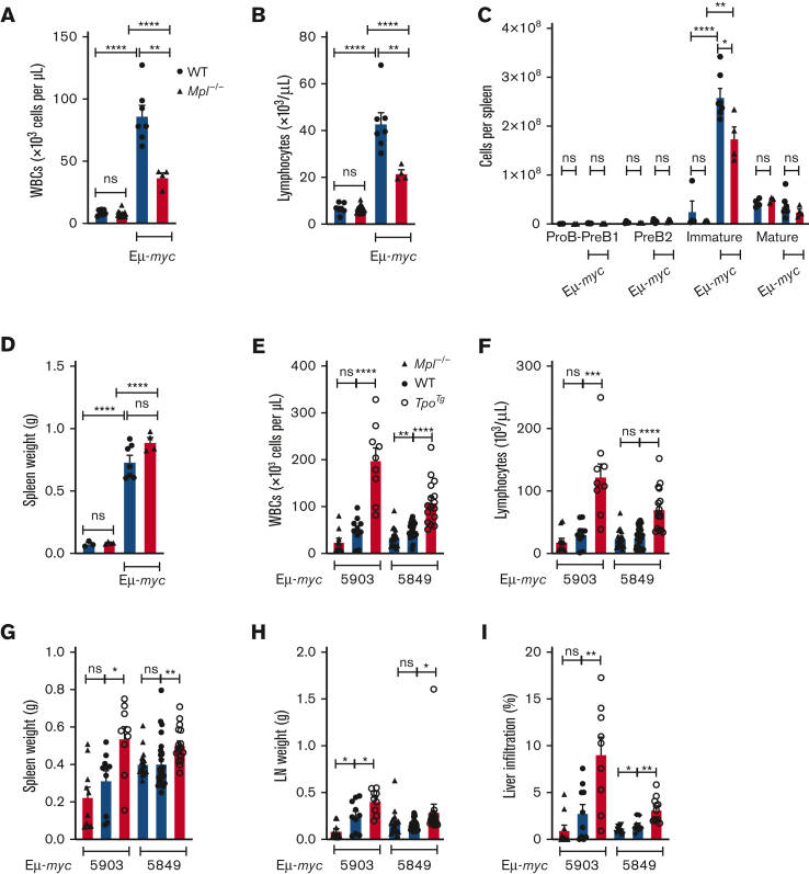
Increased lymphoma progression in TpoTg mice. (A) WBC and (B) lymphocyte counts. (C) B-cell numbers in the spleen and (D) spleen weight in WT and Mpl−/− mice 13 days after Eμ-myc 166 IV transplantation (106 P1 cells per mouse). n = 4 to 7 mice per genotype. Each symbol represents an individual mouse. Mean ± SEM. Student unpaired t test. (E) WBC and (F) lymphocyte counts, (G) spleen weight, (H) total lymph node (LN) weight, and (I) percentage liver infiltration in WT C57BL/6, Mpl−/−, and TpoTg mice 23 days after Eμ-myc 5903 IV transplantation and 17 days after Eμ-myc 5849 IV transplantation (10 000 P1 cells per mouse). 5903; n = 9 to 10 mice per genotype. 5849; WT (n = 27), Mpl−/− (n = 18), and TpoTg (n = 16) mice. Percentage liver infiltration was assessed in 11 to 12 mice per genotype. Each symbol represents an individual mouse. Mean ± SEM. Student unpaired t test, ∗P < .05, ∗∗P < .005, ∗∗∗P < .001, ∗∗∗∗P < .0001.

