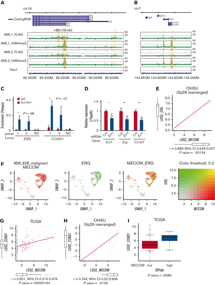
Figure 3.
ERG and CCND1 are targets of EVI1 in Evi1high AML cells. (A-B) Illustration of anti-FLAG and anti-H3K4me3 ChIP-seq results for FLAG-tagged EVI1 in the murine Erg locus around the alternative transcription start site (A) and the murine Ccnd1 promoter region (B). Yellow bars represent significantly enriched regions. (C) ChIP-qPCR analysis using anti-EVI1 antibody and HNT-34 AML cells showing the binding of EVI1 to the corresponding human regions identified in the ChIP-seq of the murine EVI1-AML samples. Neighborhood sequences without enrichment in the ChIP-seq were used as a negative control. (D) qPCR showing the relative expression of Evi1, Erg, and Ccnd1 in EVI1-AML cells expressing shRNA against Evi1. (E) Gene expression correlation analysis between MECOM and ERG in Oregon Health Sciences University (OHSU) AML cohorts with 3q26 abnormalities. (F) A 2-color dot plot showing coexpression of MECOM and ERG in the scRNA-seq data from a patient with AML (AML328). (G) Gene expression correlation analysis between MECOM and CCND1 in The Cancer Genome Atlas (TCGA) AML cohorts. (H) Gene expression correlation analysis between MECOM and CCND1 in OHSU AML cohorts with 3q26 abnormalities. (I) A box plot showing CCND1 expression in TCGA EVI1+ and EVI1− AML cohorts. Mean ± SD. ∗P < .05, ∗∗P < .01.

