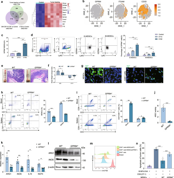
Fig. 1.
GPR84 deficiency block MDSCs immunosuppression and prevent esophageal cancer. a Venn analysis showed the upregulatory molecule in tumor-associated MDSCs and the top ten genes in esophageal cancer were shown in heatmap. b t-Distributed stochastic neighbor embedding (t-SNE) visualization of GPR84, ITGAM, and CD3D. c Comparison analysis of GPR84+ MDSC frequency in esophageal tumors in normal control (Control, n = 3), early stage (D113, n = 3) and late stage of esophageal cancer (D162, n = 9) after 4-NQO stimulation by flow cytometry. d The expression of GPR84 on M-MDSCs (Control, n = 3; D113, n = 3; D162, n = 6) and G-MDSCs (Control, n = 3; D113, n = 3; D162, n = 6) were performed in late-stage esophageal cancer (D162) by flow cytometry. e Gross and microscopic specimens of 4-NQO induced esophageal cancer model in WT and GPR84−/− mice. f Body weight was measured on days 81 and 117 after 4-NQO stimulation and the change index was calculated to determine the progression of esophageal cancer in WT and GPR84-/-mice. g Immunofluorescence analysis demonstrates the infiltration of MDSCs and CD8+ T cells in esophageal cancer tissues. h, i Flow cytometry has been performed to analyze the accumulation of MDSCs and CD8+ T cells in blood and spleen. j, k MDSCs was purified using isolation kit (Miltenyi); qRT-PCR and western blotting have been performed to investigate the different expression of immunosuppressive molecule in MDSCs between WT and GPR84−/− mice. l ARG 1 activity in MDSCs from WT and GPR84−/− mice was detected by Arginase Activity Assay Kit. m Coincubation system was performed to investigate the immunosuppression of MDSCs on CD8+ T cells proliferation. n The antitumor activity of CD8+ T cells cocultured with MDSCs from WT and GPR84−/− esophageal cancer mice were analyzed using flow cytometry. Data are represented as mean ± SEM. *p < 0.05, **p < 0.01, ***p < 0.001

