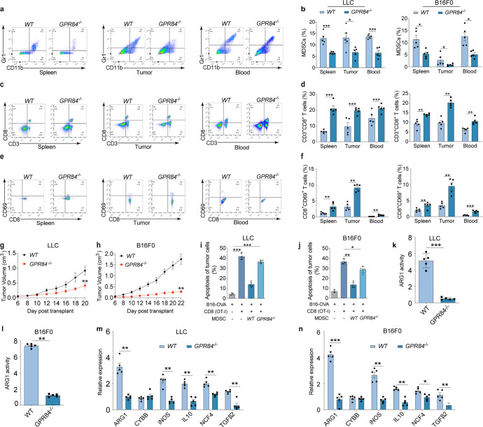
Fig. 2.
GPR84 contributes to lung cancer and melanoma progression dependent on MDSCs immunosuppression. Percentages of MDSCs (a, b), CD3+CD8+ T cells (c, d) and CD8+CD69+ T cells (e, f) in spleen, tumor tissue, and peripheral blood of LLC-cell and B16F0-cell-injected WT and GPR84−/− mice were analyzed by flow cytometry. g, h Tumor volumes of WT and GPR84−/− mice were measured after injection of LLC cells and B16F0 cells. i, j The specific antitumor activity of CD8+ T cells cocultured with MDSCs from WT and GPR84−/− tumor-bearing mice was analyzed using flow cytometry. k, l Arginase-1 activity in MDSCs from WT and GPR84−/− mice given LLC cells and B16F0 cells injection were analyzed. m, n The relative expression of immunosuppressive molecules (ARG 1, CYBB, iNOS, IL-10, NCF4, and TGF-β2) in MDSCs from WT and GPR84−/− mice were analyzed using qPCR. Data are represented as mean ± SEM. *p < 0.05, **p < 0.01, ***p < 0.001

