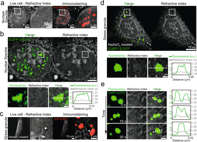
Fig. 2. Nuclear speckles and stress granules are low-density condensates.
a (Left) RI images of an intact live U2OS cell. (Right) Combined RI and immunofluorescence images of the same cell after fixation. Anti-SRSF2 is used to target nuclear speckles. b (Top) Combined RI and fluorescence images of a live U2OS cell expressing EGFP-SRSF2. (Bottom left) Zoomed-in images of a single nuclear speckle. (Bottom right) Intensity profile plot for normalized fluorescence intensities of EGFP-SRSF2 (green line) and RIs (gray line) along the white dashed line. c (Left) RI images of a live U2OS cell treated with 500 μM sodium arsenite. (Right) Combined RI and immunofluorescence images of the same cell after fixation. Anti-G3BP1 is used to target stress granules. d (Top) Combined RI and fluorescence images of a live U2OS cell expressing G3BP1-EGFP after the treatment of 500 μM sodium arsenite. (Bottom left) Zoomed-in images of a single stress granule. (Bottom right) Intensity profile plot for normalized fluorescence intensities of G3BP1-EGFP (green line) and RIs (gray line) along the white dashed line. e (Left) Time-lapse images of a fusion event between two stress granules. (Right) Intensity profile plots for normalized fluorescence intensities of G3BP1-EGFP (green line) and RIs (gray line) along white dashed lines. All refractive index images are adjusted to 1.337–1.37.

