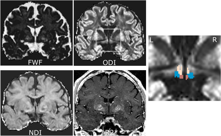
Figure 4.
Exemplary dataset in native space displaying the four used MRI parameters in coronal view (FWF: free water fraction, ODI: orientation dispersion index corrected for FWF, NDI: neurite density index corrected for FWF, R2*: effective transverse relaxation rate), as well as the ODI image zoomed in on the hypothalamus area and overlaid with the hypothalamus masks after correction for confounding white matter (beige: superior, blue: intermediate, brown: posterior subunits). The inferior subunit is not visible on this slice.

