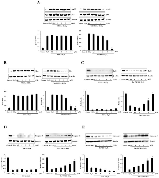
Fig. 3.
Effect of Tat-TXNL1 protein against H2O2-induced apoptotic protein expression in HT-22 cells. The cells were treated with Tat-TXNL1 (0.5-7 μM) or TXNL1 for 1 h and exposed to H2O2 (1 mM). The expression of (A) p53, (B) Bax, (C) Bcl-2, (D) Pro-caspase-9 and (E) Pro-caspase-3 were analyzed by Western blotting. The band intensity was measured by densitometry. *P < 0.05 and #P < 0.01 compared with H2O2 treated cells. The bars in the figure represent the mean ± SEM obtained from 3 independent experiments.

