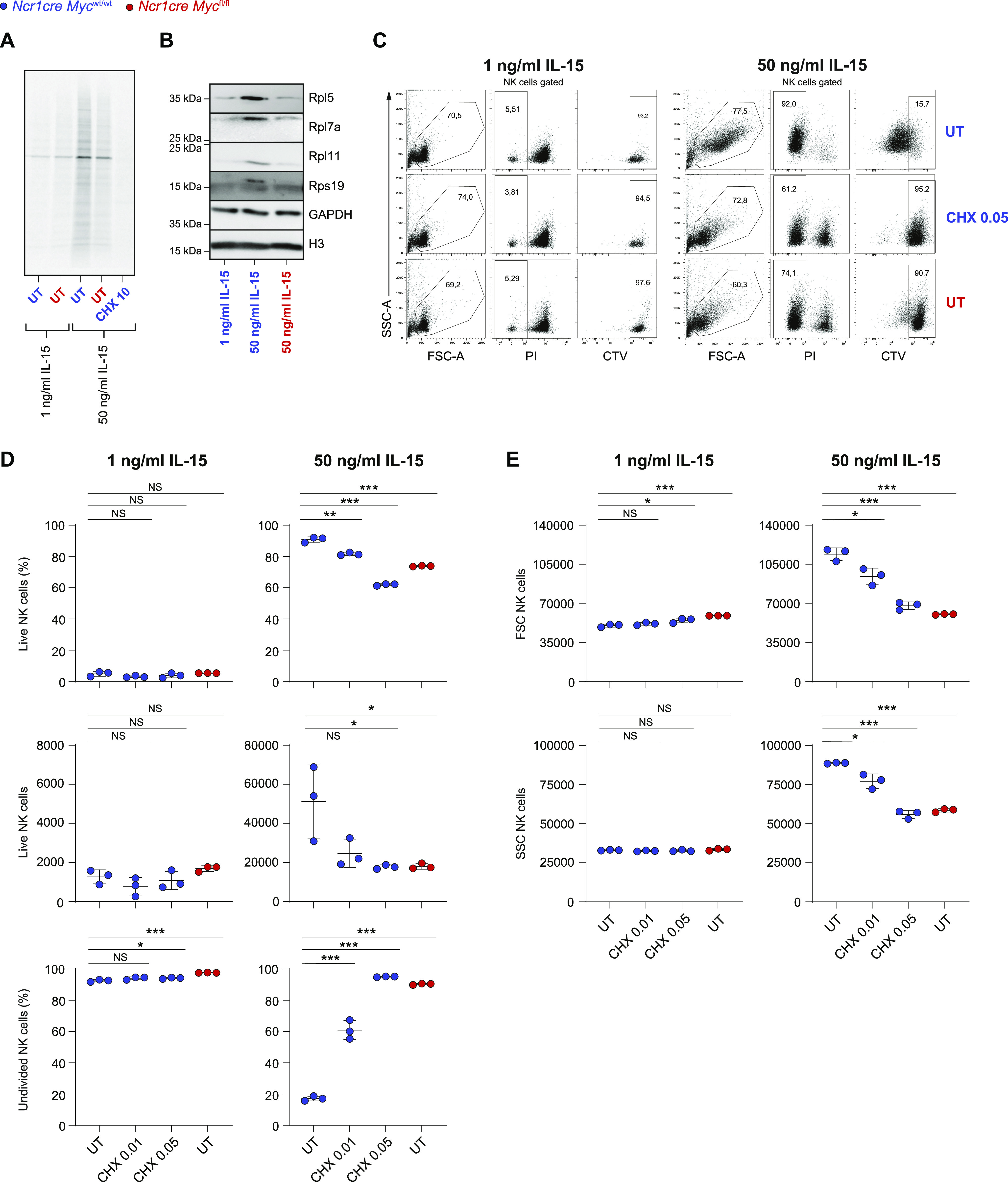
Figure 5. Full translational capacity is required in response to acute IL-15.

(A) Equal numbers of NK cells enriched from splenocytes of Ncr1cre Mycfl/fl and Ncr1cre Mycwt/wt mice were stimulated overnight with 1 or 50 ng/ml IL-15 and incorporation of 35S-radiolabeled methionine and cysteine was assessed. CHX (10 μg/ml) was added as a control where indicated. (B) CD11b+ cells (gated as NK1.1+CD11b+CD3−) were sorted from total NK cells enriched from the spleens of Ncr1cre Mycfl/fl and Ncr1cre Mycwt/wt mice and stimulated overnight with IL-15 as indicated. The protein expression levels of Rpl5, Rpl7a, Rpl11, and Rps19 were assessed by Western blotting. Gapdh and Histone H3 levels are shown as loading controls. (C, D, E) Enriched Myc-deficient or control splenic NK cells were stimulated with the indicated doses of IL-15. After an overnight period, 0.01 or 0.05 μg/ml of cycloheximide was added and the cells were further cultured for 3 d. (C) Representative flow cytometric dot plots gated on NK1.1+ cells show live NK cells (PI−) or cells division (CTV dilution). (D, E) Graphs show percentages, absolute numbers, percentages of undivided cells (D), FSC, and SSC values (E) of live NK cells. (A, B, C, D, E) Results are representative of at least two experiments. (D, E) Results show mean ± SD of technical triplicates. (D, E) Statistical comparisons are shown; *P ≤ 0.05, **P ≤ 0.01, ***P ≤ 0.001, and NS, non-significant; t test, unpaired (D, E).
