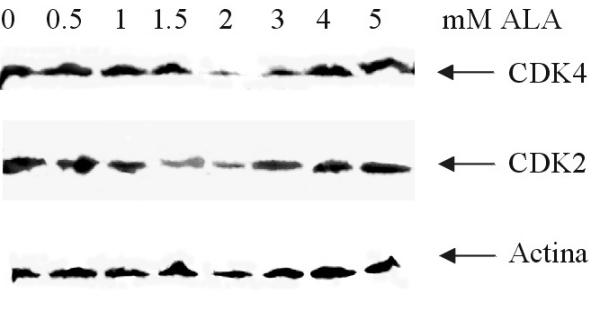
Figure 6.

Western blot analysis of CDK4 and CDK2. Proteins were isolated after treatment of cells with ALA (0–5 mM) during 24 h. Proteins were normalized to 50 μg per lane and the amount of CDK2 and CDK4 was detected using polyclonal antibodies and visualized using ECL reagents. Intensity of bands was analysed with ImageMaster.
