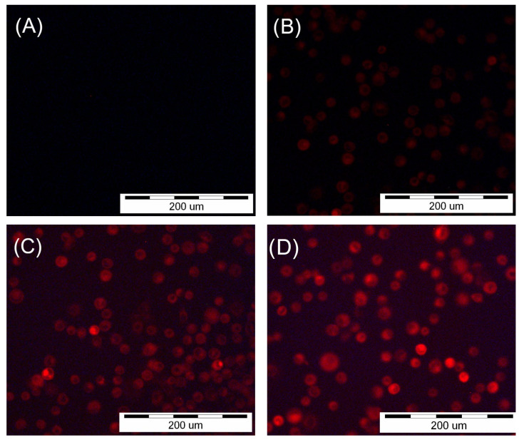
Figure 1.
Images of the SCC-25 cells were obtained using an inverted fluorescence microscope after two-hour incubation with HY. (A) No fluorescence after incubation with 0 µM HY solution. (B) Fluorescence after incubation with 0.25 µM HY solution. (C) Fluorescence after incubation with 0.5 µM HY solution. (D) Fluorescence after incubation with 1 µM HY solution.

