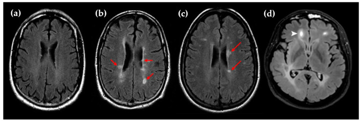
Figure 1.
(a) Representative MRI scan (T2/FLAIR) of MS patients without brain lesions; (b,c) Representative MRI scans (T2/FLAIR) of MS patients with numerous brain lesions (red arrows); (d) Representative Gd-enhanced lesion in a frontal lobe (white arrow). The images (a–c) are reproduced from Dastagir et al. with the addition of red arrows (BY-NC-ND/4.0 license) [4]. Image (d) is reproduced from Lopaisankrit et al. without changes (CC BY/4.0 license) [5].

