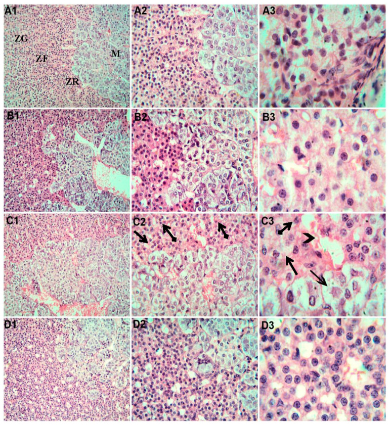
Figure 4.
Representative adrenal gland sections photomicrographs. (A1,A2,A3) Control rats demonstrating the normal structure of the zona glomerulosa (ZG), zona fasciculata (ZF), zona reticularis (ZR), and medulla (M). (B1,B2,B3). Rats that received Mg and showing normal histological architecture and arrangement of cells within the adrenal cortex and medulla. (C1,C2,C3) Rats exposed to SiNPs showing disorganized cell cords interspersed with distended blood sinusoids (arrow heads) and abnormal cortical cells, which show vacuolated cytoplasm (arrows) and pyknotic nuclei (tailed arrows). H&E. (A1,B1,C1,D1) ×20 magnification. (A2,B2,C2,D2) ×40 magnification. (A3,B3,C3,D3) ×100 magnification.

