Figure 5.
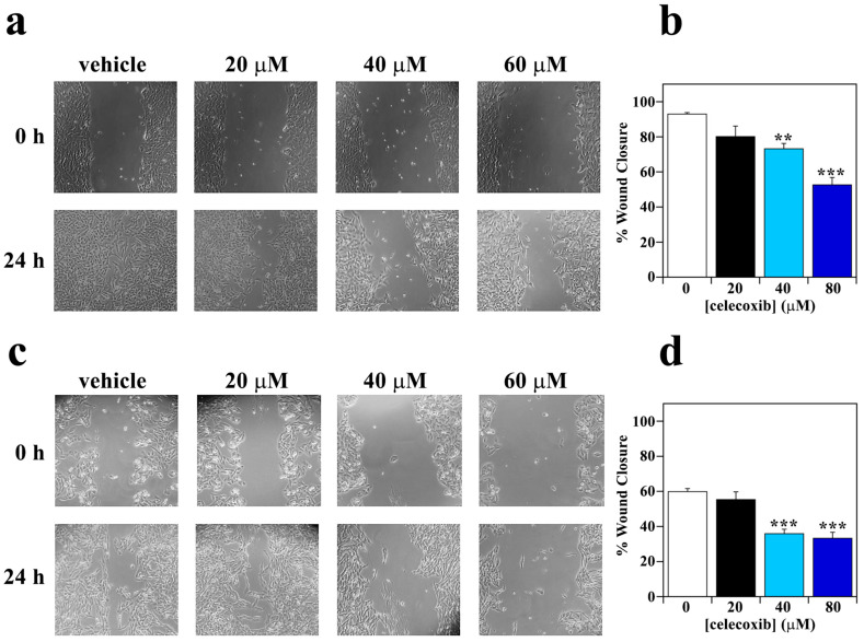
Effect of celecoxib on migratory capability of melanoma cells. (a) A2058 cells and (c) SAN cells were wounded with a pipette tip and photographed immediately after the wounding (0 h) and after 24 h of treatment with vehicle alone (0.5% (v/v) DMSO) or 20, 40 or 60 µM celecoxib. Quantification of migration capacity of (b) A2058 cells and (d) SAN cells incubated for 24 h with vehicle alone (0.5% (v/v) DMSO) (white bar), 20 µM celecoxib (black bar), 40 µM celecoxib (cyan bar), or 60 µM celecoxib (blue bar). Eight fields per scratch were measured to achieve an objective evaluation. Data are expressed as percentage of wound closure after 24 h of treatment with respect to 0 h and reported as the mean of two independent experiments ± SE. ** p < 0.01; *** p < 0.0001 compared to control cells.

