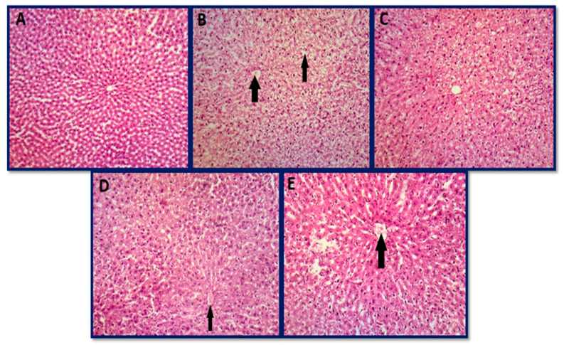
Figure 10.
Histopathological photomicrographs of the liver: (A) group A (control group), (B) group B (aflatoxicated group) showed swelling of hepatocytes with portal infiltration with neutrophils, (C) group C (toxin + 20 µg AgNPs) normal architecture, (D) group D (toxin + 50 µg AgNPs) showed mild swelling of hepatocytes with distorted hepatic cords as compared to the toxin group, and (E) group E (toxin + 70 µg AgNPs) showed mild swelling of hepatocytes and leukocyte infiltration in hepatic cords (hematoxylin and eosin 10×).

