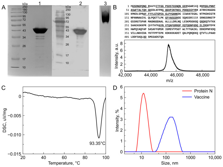
Figure 1.
Biochemical characterization of recombinant SARS-CoV-2 N protein. (A) SDS-PAGE and Western blotting confirm the molecular weight and recognition by N-specific antibodies. Native PAGE shows homogeneous distribution of recombinant N as one species. Lanes: 1—SDS-PAGE of recombinant N protein solution; 2—Western blot of recombinant N protein solution; 3—native PAGE of recombinant N-protein solution. Uncropped photo of Western Blot membrane is presented in Figure S2. (B) MALDI-MS confirms the predicted molecular weight of monomeric recombinant nucleocapsid protein and 96.7% of sequence coverage was achieved. Confirmed sequence is marked in bold. Peptides subjected to MS/MS analysis are underlined. (C) Differential scanning calorimetry thermograms of 0.2 mg/mL N-protein samples show no endothermic peaks corresponding to protein globule melting but show a distinct exothermic peak at 93 °C corresponding to oligomer particles melting. (D) Nucleocapsid protein forms oligomeric structures with a hydrodynamic diameter of approximately 13 nm (red line) which corresponds to a Mw of approximately 200 kDa suggesting the formation of a tetramer. The emulsion formed by squalane, (D,L)-α-tocopherol and polysorbate 80 exposes the protein on oil in water drops with a size of approximately 150 nm in diameter (blue line).

