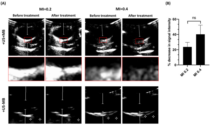
Figure 4.
(A) Ultrasound contrast images from porcine eyes treated with UMSB (+US+MB) or ultrasound only (+US−MB) at MI 0.2 and 0.4. Red boxes indicate enlargement of the focal area of the probe. Contrast signal was minimally (MI 0.2) and strongly (MI 0.4) decreased after USMB treatment. Distance between the two crosses is 1 cm. (B) Percentage of decrease in signal intensity of microbubbles within the PW Doppler cursor area after treatment with USMB at MI 0.2 and 0.4 (n = 4), ns: not significant.

