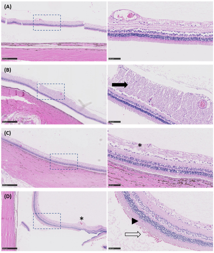
Figure 9.
Microphotographs of H&E-stained sections from eyes exposed to different USMB conditions. Images on the right (20× magnification, scale bar: 100 μm) are high magnification views of dashed-line boxes shown on the left (5× magnification, scale bar: 500 μm). (A) Untreated eye (i.e., no USMB) with no signs of retinal damage. (B) Eye treated with USMB at MI 0.2 showing some edematous changes (black arrow) in the proximity of the optic nerve. (C) In the eye treated with USMB at MI 0.4, focal degenerative vacuolization with fibrinous exudate was observed (asterisk). (D) The most pronounced damage was seen in the eye treated with USMB at MI 0.8. Focal degenerative vacuolization with fibrinous exudate was seen around the retinal blood vessel (asterisk), erythrocytes were present in the subretinal space (hollow arrow) and loss of cell boundaries was seen in the photoreceptor layer (arrowhead).

