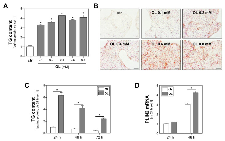
Figure 1.
Effect of oleate treatment on lipid accumulation in Calu-3 cells. (A,B) Calu-3 cells were incubated with increasing oleate (OL) concentrations for 24 h. Cells treated with FFA-free BSA served as controls (ctr). (A) Triglyceride (TG) content normalized to cellular protein content (*: p < 0.05 compared to ctr). (B) Oil Red O staining; scale bars 50 µm. (C,D) Calu-3 cells were incubated with 0.6 mM OL or FFA-free BSA (ctr) for different time intervals, as indicated. (C) Triglyceride (TG) content normalized to cellular protein content and (D) PLIN2 mRNA levels analyzed by quantitative RT-PCR (*: p < 0.05).

