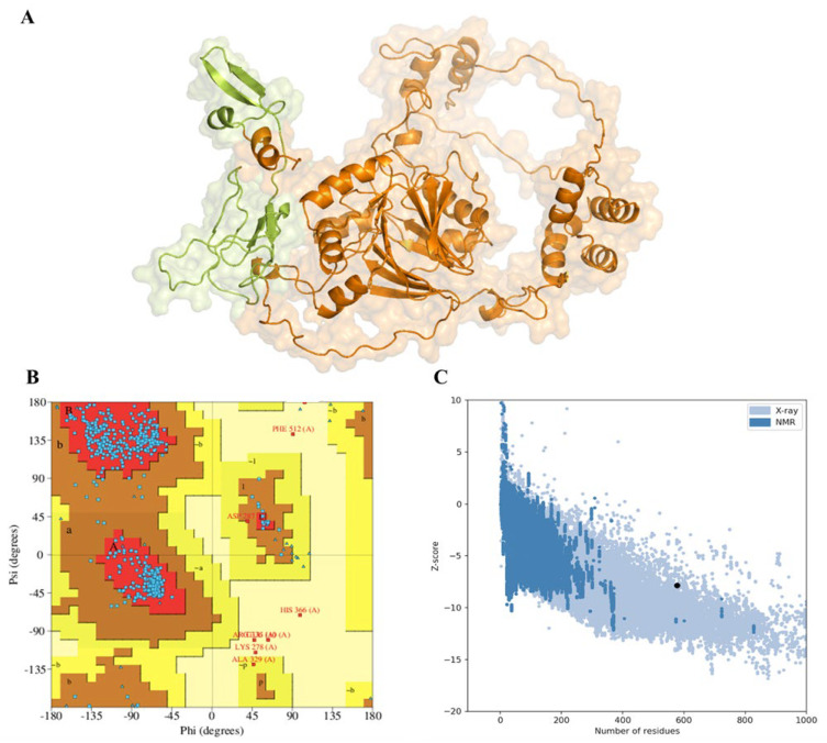
Figure 2.
The predicted tertiary structure of bovine CMAH protein and its validation. (A) Tertiary structure of CMAH. Lime colour indicates iron-sulphur domain (14–112). (B) Assessment and validation of the protein. Ramachandran plot depicted that 92.6% of the amino acids are located in the most favoured region with a total of 462 residues (A, B, L), 5.8% in the additional allowed region with a total of 29 residues (a, b, l, p), 0.4% in the generously allowed region with two residues (~a, ~b, ~l, ~p), and 1.2% in the disallowed region with six residues. (C) Prosa plot with a z-score of −7.86. The black dot represents the position of the bCMAH structure compared with the standard X-ray crystallography parameters for proteins of a similar size.

