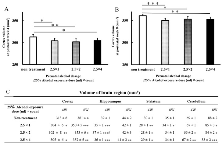
Figure 3.
Graphs of postnatal cerebral volume at 4 weeks (A) and 8 weeks (B). Brain volumes for each brain area at 4 and 8 weeks postnatally: cortex, hippocampus, striatum, and cerebellum (C). In the FASD group that received alcohol, there was a larger decrease in brain volume in each brain region compared with that of the non-treated group. Brain volume reduction of the brain regions occurred at 4 and 8 weeks of age. Non-treatment = normal control group (n = 11); 2.5 × 1 = one dose of 2.5 mL of 25% alcohol (n = 11); 2.5 × 2 = two doses of 2.5 mL of 25% alcohol (n = 7); 2.5 × 4 = four doses of 2.5 mL of 25% alcohol (n = 11). Between-group comparisons were analyzed by one-way ANOVA with Tukey’s multiple comparison tests using Prism 9. The results showed no significant difference between the two-dose group (2.5 × 2) and the four-dose group (2.5 × 4). As compared with the non-treatment group: * p < 0.05; ** p < 0.01; *** p < 0.001. As with the one-dose FASD group (2.5 × 1): # p < 0.05.

