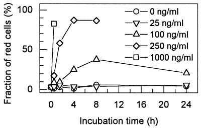
FIG. 1.
Killing by T4 lysozyme of B. subtilis 168 cells in 0.3× PBS with 50% glycerol followed by fluorescence microscopy. The suspensions were incubated at various T4 lysozyme concentrations for up to 24 h and stained with the BacLight bacterial viability fluorescence dyes (see Materials and Methods). The fraction of red (dead) cells is given for each time point. Several independent determinations of the fraction of red cells during a 30-min interval with different T4 lysozyme concentrations gave the following data (means with standard deviations): 100 ng/ml, 10.4% ± 5.2% (n = 4); 250 ng/ml, 17.3% ± 6.9% (n = 3); 1,000 ng/ml, 70.5% ± 12.9% (n = 4).

