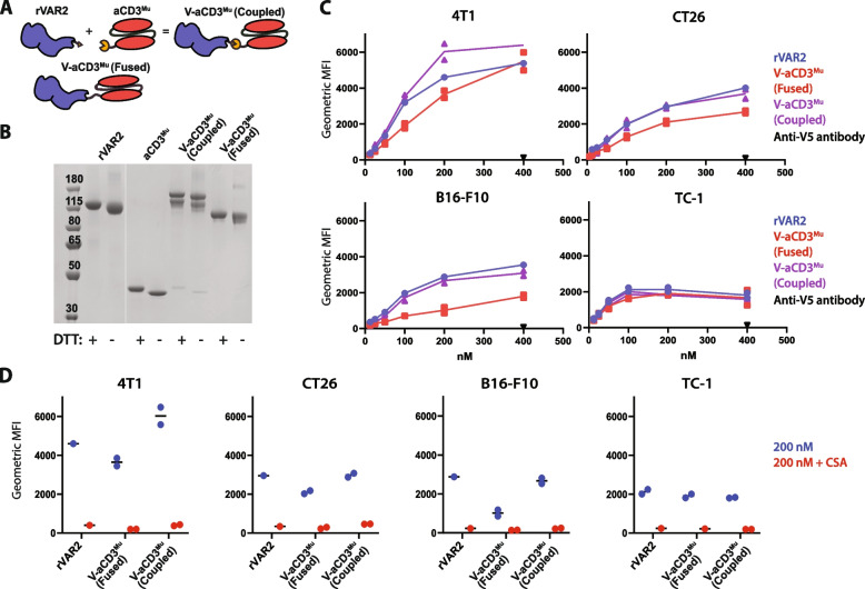
Fig. 1.
rVAR2 coupled to aCD3Mu retains cancer cell binding. A Schematic figure of rVAR2 and aCD3Mu conjugated through the SpyTag/SpyCatcher system into one protein (V-aCD3Mu (coupled)) very similar to the genetically fused V-aCD3Mu. B SDS-PAGE of rVAR2, aCD3Mu, V-aCD3Mu (coupled), and V-aCD3Mu (fused). C Flow cytometry showing binding of rVAR2, V-aCD3Mu (coupled), and V-aCD3Mu (fused) to the indicated cancer cell lines, including the detection antibody anti-V5 as a control. D Flow cytometry showing binding of 200 nM of indicated protein with and without soluble chondroitin sulfate A (CSA) added in excess. Each dot represents one data point. TC-1 binding of V-aCD3Mu (coupled) and V-aCD3Mu (fused) were evaluated in individual experiments and values were normalized relative to rVAR2 binding ((V-aCD3Mu (coupled)/rVAR2_1) * VAR2_2). Data are representative of either two (B16-F10 and TC-1) or four (4T1 and CT26) individual experiments

