Fig. 2.
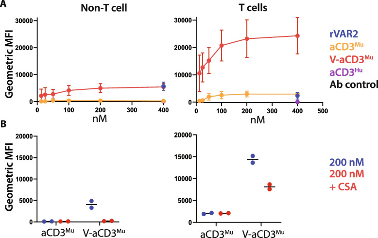
aCD3Mu retains T cell binding when linked to rVAR2. A Flow cytometry on protein binding to non-T cells (CD4-CD8-) and T cells (CD4 + /CD8 + /CD4 + CD8 +) from splenocytes and white blood cells. An anti-human anti-CD3 (aCD3Hu) and an antibody control (anti-CD8, anti-CD4, and the secondary antibody anti-penta-HIS) were included as controls. Means and standard deviations are displayed. B CSA inhibition of protein binding to non-T cell splenocytes/white blood cells (left panel) and T cells (right panel). Each dot represents one data point. Data in this figure is compiled from three separate experiments. Note that the fluorescent signals cannot be directly compared as V-aCD3Mu has two penta-HIS tags while aCD3Mu, rVAR2, and aCD3Hu only have one

